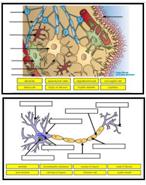
The labeling focuses on the neuron and supporting neuroglia (or glial) cells. These cells provide provide physical and metabolic support to neurons. Neuroglia cells are different from nerve cells in that they do not participate directly in synaptic interactions. Students can also label a nerve cell and color neuroglia cells using paper handouts.
Central nervous system or CNS brain organ structure outline diagram. Labeled educational scheme with cerebrum, brainstem and cereb… | Anatomie, Biologie, Neurologie
Question: Art-labeling Activity: Neuron structure9 of 9labels to the appropriate location in the figure. Art-labeling Activity: Neuron structure. 9 of 9. labels to the appropriate location in the figure. Here’s the best way to solve it. Powered by Chegg AI. Step 1. View the full answer.

Source Image: pinterest.com
Download Image
Study with Quizlet and memorize flashcards containing terms like Label the following structural components of a neuron., Correctly label the cells of the central nervous system on the diagram., Correctly match the labels to the following parts of a cholinergic synapse and more.

Source Image: pinterest.com
Download Image
Neurons May 7, 2022The neuron is one of two basic types of cells in the nervous system, the other type being the glial cell. Figure \ (\PageIndex 1\): Interneurons of Adult Visual Cortex. Neurons, also called nerve cells, are electrically excitable cells that are the main functional units of the nervous system. Their function is to transmit nerve impulses.

Source Image: enchantedlearning.com
Download Image
Art Labeling Activity Neuron Structure
May 7, 2022The neuron is one of two basic types of cells in the nervous system, the other type being the glial cell. Figure \ (\PageIndex 1\): Interneurons of Adult Visual Cortex. Neurons, also called nerve cells, are electrically excitable cells that are the main functional units of the nervous system. Their function is to transmit nerve impulses. Jun 14, 2023The fluorescent styryl dye FM 1-43 was previously shown to label sensory neurons. Surprisingly, we find that the vast majority of FM 1-43 somatosensory neuron labeling in mice in vivo is dependent on PIEZO2 activity within the peripheral nerve endings. We illustrate the potential of FM 1-43 by using it to identify novel PIEZO2-expressing
Label Neuron Anatomy Printout – EnchantedLearning.com
Cortical neuronal pathways travel outside of the spinal cord to the cerebral cortex and are slower than spinal neuronal pathways. Study with Quizlet and memorize flashcards containing terms like Art-labeling Activity: Nerve fibers, Art-labeling Activity: Structure of a typical motor neuron (2 of 2), Which factors contribute to increasing the Neuron Labeling
Source Image: biologycorner.com
Download Image
Printable Worksheet: Neuron Diagram Cortical neuronal pathways travel outside of the spinal cord to the cerebral cortex and are slower than spinal neuronal pathways. Study with Quizlet and memorize flashcards containing terms like Art-labeling Activity: Nerve fibers, Art-labeling Activity: Structure of a typical motor neuron (2 of 2), Which factors contribute to increasing the

Source Image: brainframe-kids.com
Download Image
Central nervous system or CNS brain organ structure outline diagram. Labeled educational scheme with cerebrum, brainstem and cereb… | Anatomie, Biologie, Neurologie The labeling focuses on the neuron and supporting neuroglia (or glial) cells. These cells provide provide physical and metabolic support to neurons. Neuroglia cells are different from nerve cells in that they do not participate directly in synaptic interactions. Students can also label a nerve cell and color neuroglia cells using paper handouts.

Source Image: pinterest.com
Download Image
Neurons Study with Quizlet and memorize flashcards containing terms like Label the following structural components of a neuron., Correctly label the cells of the central nervous system on the diagram., Correctly match the labels to the following parts of a cholinergic synapse and more.

Source Image: pinterest.com
Download Image
Neuron Anatomy Lesson Neuron Cell Labeling & Functions Science Worksheet. by. TechCheck Lessons. 4.5. (10) $0.99. Zip. This resource contains 2 worksheets for students to label the common/major parts of neuron cells and complete a chart detailing the major functions of each part. Answer key included.This resource can be used as an introduction to new material or a

Source Image: pinterest.com
Download Image
Brain And Neuron Drawings | Brain art, Medical drawings, Human anatomy art May 7, 2022The neuron is one of two basic types of cells in the nervous system, the other type being the glial cell. Figure \ (\PageIndex 1\): Interneurons of Adult Visual Cortex. Neurons, also called nerve cells, are electrically excitable cells that are the main functional units of the nervous system. Their function is to transmit nerve impulses.

Source Image: pinterest.com
Download Image
Premium Vector | Neuron anatomy of human cell line art vector and illustration design neuron anatomy and human cell Jun 14, 2023The fluorescent styryl dye FM 1-43 was previously shown to label sensory neurons. Surprisingly, we find that the vast majority of FM 1-43 somatosensory neuron labeling in mice in vivo is dependent on PIEZO2 activity within the peripheral nerve endings. We illustrate the potential of FM 1-43 by using it to identify novel PIEZO2-expressing

Source Image: freepik.com
Download Image
Printable Worksheet: Neuron Diagram
Premium Vector | Neuron anatomy of human cell line art vector and illustration design neuron anatomy and human cell Question: Art-labeling Activity: Neuron structure9 of 9labels to the appropriate location in the figure. Art-labeling Activity: Neuron structure. 9 of 9. labels to the appropriate location in the figure. Here’s the best way to solve it. Powered by Chegg AI. Step 1. View the full answer.
Neurons Brain And Neuron Drawings | Brain art, Medical drawings, Human anatomy art Neuron Cell Labeling & Functions Science Worksheet. by. TechCheck Lessons. 4.5. (10) $0.99. Zip. This resource contains 2 worksheets for students to label the common/major parts of neuron cells and complete a chart detailing the major functions of each part. Answer key included.This resource can be used as an introduction to new material or a